Division of Integrative Systems and DesignHKUST
← All research stories

Solve / Create / 2023

MaSoChains

Small during insertion. Functional after deployment.

THE CENTRAL QUESTION

How can a functional tool pass through a narrow catheter and become much larger at the treatment site?

Minimally invasive tools face a basic geometric conflict. They must be small enough to pass through a catheter or natural opening, yet large and structured enough to image, grip, steer or perform a procedure. Making every component smaller eventually weakens the tool and removes useful functions. MaSoChains take a different route: deliver a simple chain, then let it assemble into the working device inside the body.

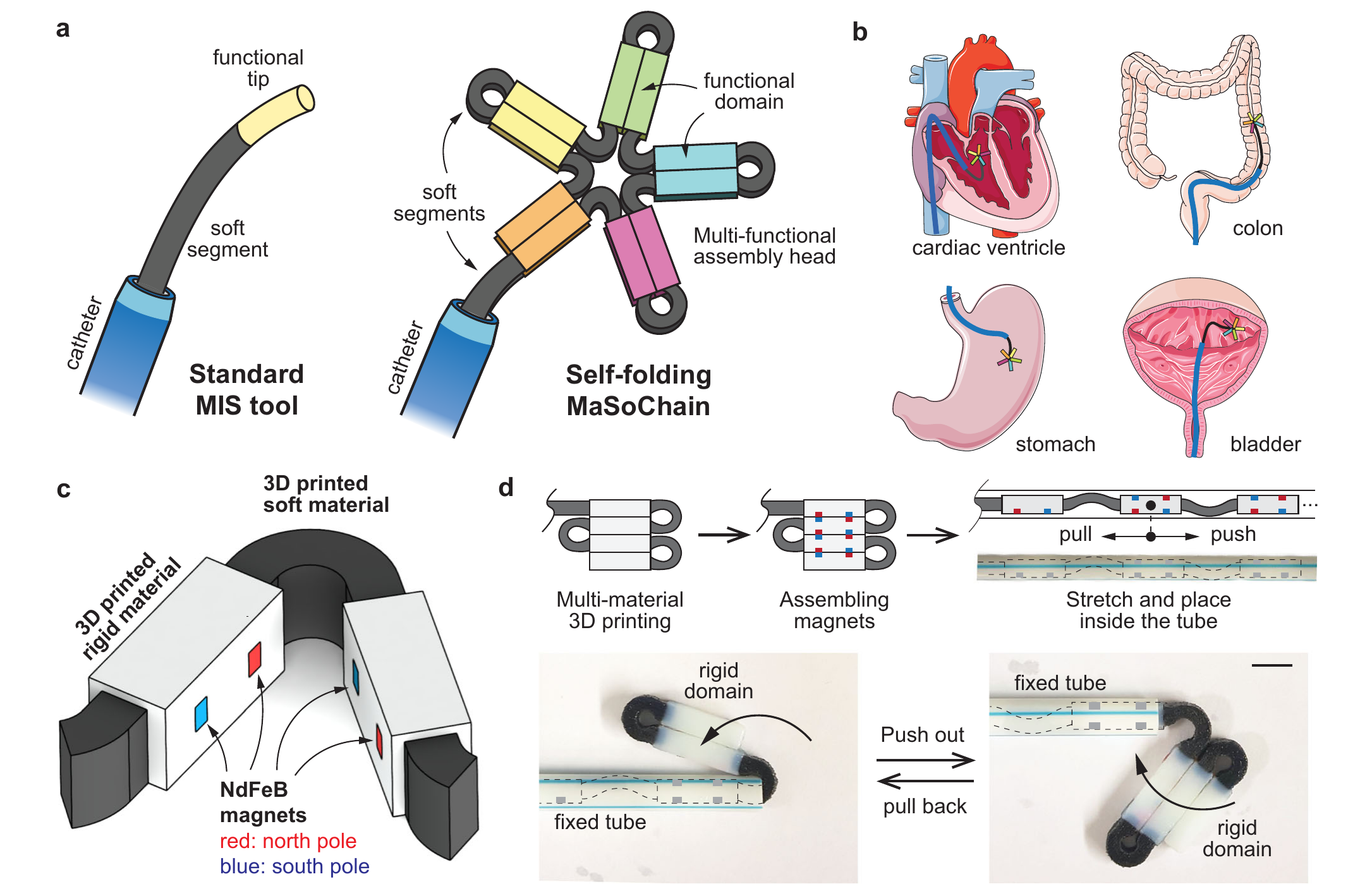
A programmed rigid-soft chain folds after catheter delivery and can be retrieved again.
A programmed rigid-soft chain folds after catheter delivery and can be retrieved again. Cropped from Fig. 1 of the linked paper.
01

Separating delivery shape from working shape

Instead of treating the tool as one fixed object, the design gives it two useful states. Inside the catheter, the chain is straight and compact. After extrusion, it folds into a larger three-dimensional arrangement. Retraction reverses the process, so deployment does not sacrifice recoverability.

02

How the chain folds and locks

Rigid domains are connected by elastic soft links that store bending energy while constrained in the catheter. As each segment exits, the stored energy bends the joint toward its programmed angle. Small embedded magnets then pull neighbouring rigid domains together and latch the final configuration without an additional locking mechanism.

03

Testing the mechanism before claiming a device

The work measured folding dynamics, elastic forces and magnetic holding forces across different geometries. These measurements showed when a joint folds reliably, when magnetic attraction is strong enough to close it and how catheter friction or material viscoelasticity can disturb the sequence.

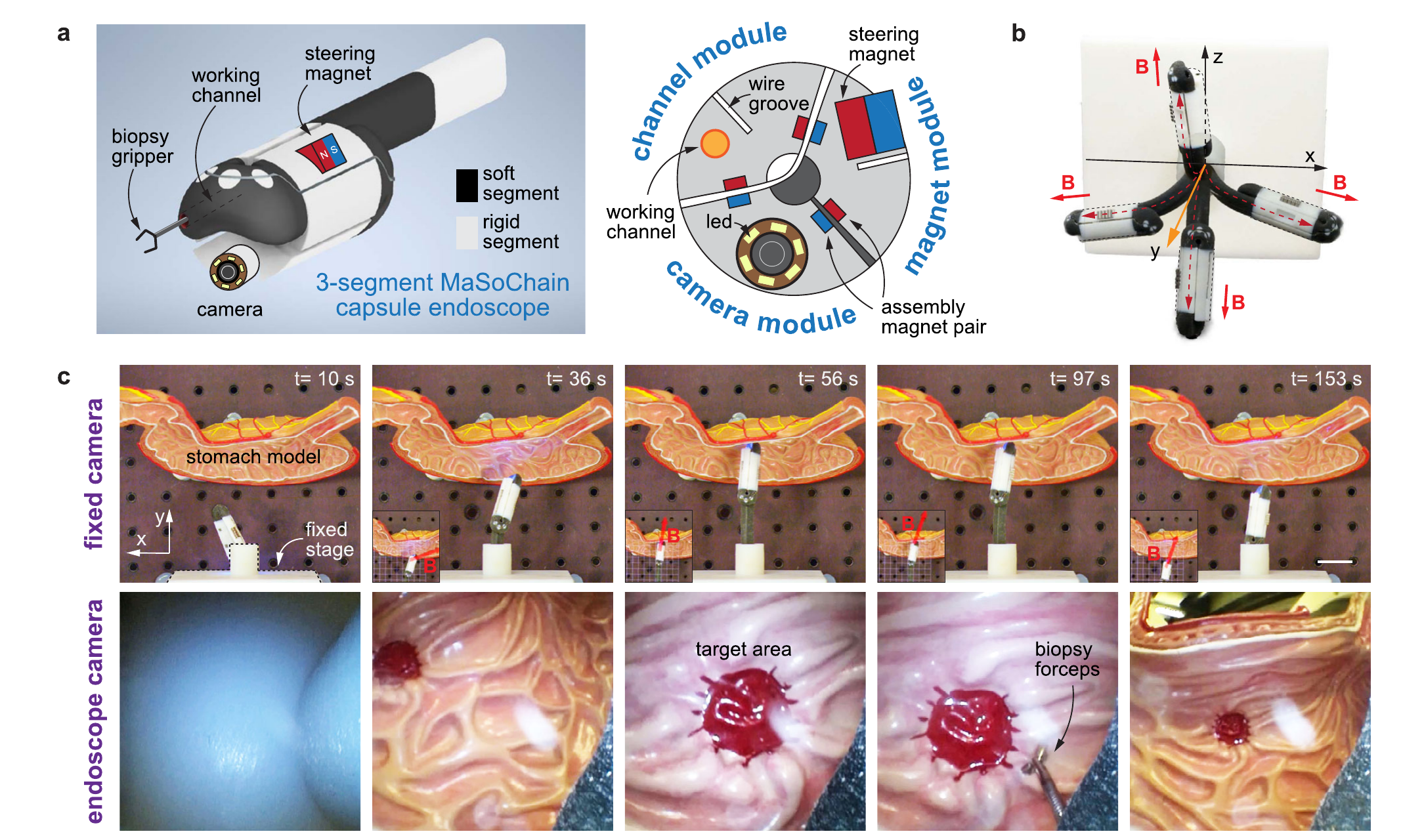
A capsule-endoscope demonstrator combines imaging, navigation and biopsy in a stomach model.
A capsule-endoscope demonstrator combines imaging, navigation and biopsy in a stomach model. Cropped from Fig. 9 of the linked paper.
04

A complete intervention sequence

The capstone demonstrator joined three modules: a camera, a magnetic steering element and a working channel for a biopsy gripper. In a stomach model, the chain assembled after delivery, was guided toward a target, supported visual inspection and biopsy, and then folded back into the catheter for removal.

05

What remains before clinical use

The demonstrations are preclinical and benchtop. The current 3D-printed elastomer does not provide enough elastic force when scaled below roughly one millimetre, and real procedures add friction, uncertain anatomy, sterilization and reliability requirements. The transferable idea is to assemble function at the destination rather than allowing the access diameter to define the entire device.